A microscope (in Greek: mikros, “small’ and, skopein, “to look” or “see”) is an instrument to see objects too small for the naked eye. The science of investigating small objects using such an instrument is called microscopy. In Physiology. The compound microscope (a light microscope consisting of multiple lenses) is the tool that can be used for many purposes e.g. studying the morphology of various cells and different types of cell counts. It is necessary to understand the physical principles associated with the microscope to obtain the best performance and results.
Types of Microscopes:
Brightfield (Compound) microscope
2. Darkfield microscope
Phase-contrast microscope
4.. Electron microscope (EM)
Transmission electron microscope (TEM)
Physical terms
Magnification:
The perceived increase in the size of an object upon viewing through a microscope.
Total magnification obtained with a compound microscope is equal to the product of the magnification supplied by the ocular lens and that supplied by the objective lenses e.g.10X x 100X=1000X
Usually. A magnification of 1000X is the best that compound microscope can give us.
Resolution [resolving power]: The ability of a microscope to present a magnified image of closely spaced points as separate points.
Focal length: The focal length of an optical system is a measure of how strongly the system converges (focuses) or diverges light.
Numerical aperture (In optics): The numerical aperture (NA) of an optical system Is a dimensionless number that characterizes the range of angles over which the system can accept or emit light
Refraction and refractory Index: The refraction of an optical system is the ability to change the direction of ray of light. The degree to which the ray of light is bent is known as refractory index.
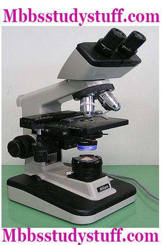
COMPOUND MICROSCOPE:
It is the most common type of microscope use. It is called brightfield because when there is no specimen, the light observed is the brightest.
A compound microscope are in use in such away that the image observes is the product of light that passes through the specimen and lens system.
3, Thus. the background is brighter than the Specimen.
Because brightfield microscopy relies solely on refraction, most specimens are difficult to see without staining.
STRUCTURAL PARTS
Scientists divides the compound microscope typically into the following structural components:
The support system
The illumination system:
III. The magnification system
lV. The adjustment system
The Support System
Base: The base of a compound microscope helps in supporting the system.
Arm: The arm acts as a connector between the base and the head of the compound microscope.
Body Tube: This part supports the eyepiece and objectives Mechanical tube length is the distance from the top of the eyepiece tube to bottom of the objective holder.
Stage: This is the platform that supports the specimen (which is typically mounted on a glass slide). Stage clips hold the slide in place.
Nosepiece (objective changer): A rotating device to which objectives (usually four) attaches.
The Illumination system:
1.Light Source: This helps in illuminating and visualizing the specimen kept on the stage. The light source may be internal (i.e electric lamp in modern microscope) or external (i.e. mirror in older versions).
2. Aperture iris diaphragm: This device is part of the substage condenser. It serves to control the angle of the cone of light emerging from the top of the condenser. It improves the resolution of the image.
3. Condenser: It is designed to control the concentration of light from the lamp onto the specimen. The optical elements can introduce a variety of aberrations which the condenser correctes it.
III. Magnification System:
Objective lens: This is either of the tour lenses attached to the nosepiece. Four different objective lenses are used in order to impart varying degrees of magnification. Conventionally used objective lenses are 4X.10X, 40X (the high power lens). Md 100X (the oil immersion lens used for differential leukocyte count).
Eyepiece (Ocular lens): This component magnifies the ‘virtual image formed by the objective lens. The ocular lens imparts the final magnification of the image observed. It usually magnifies to the order of 1OX.
Total Magnification of the Compound Microscope:
It is the product of magnification provided by objective lens and eyepiece e.g., with an eyepiece of lOX the magnification provided by 4X objective lens is 40times;
10X Objective lens is 100 times;
High power objective lens (40X) is 400 times.
Oil immersion lens (100X) is 1000 times.
The Adjustment System:
Coarse Adjustment Knob: This control (typically a pair, one on each side) moves the stage upon down in a quick manner for gross focusing.
Fine focus knob: This control allows for precise focusing of the specimen.
Condenser locus knob: This control is used precisely adjust the vertical height at the condenser.
APPARATUS:
Compound Microscope, cedarwood oil, stained slides, blood film.
PROCEDURE:
For non-stained slides:
1.Turn on the lamp.
2.Turn the lever diaphragm of the microscope completely clockwise. Fully opening the iris.
Place the prepared slide on the microscope’s stage.
Position the stage in close proximity to the objective lens.
Look through the pair of eyepieces at the specimen present on the slide.
Using 4X objective, focus on the specimen by turning the large focus knob on the microscope until you get a clear, overall view of the specimen.
Zoom in on the tiny, intricate details of the specimen by turning the small locus knob Keep turning until the small details appear clear and sharp.
Turn the diaphragm counter-clockwise. Until the level oi light hitting the specimen is satisfactory to suit your eyes.
Rotate the nosepiece on the microscope to focus with 10Xandthenwilh40Xiorhigher magnification.
For stained slides:
Use oil immersion lens (100X) for stain slides.
Place a drop of cedarwood oil on the slide.
Dip the oil immersion lens into the drop oil.
Repeat steps 4 to 8 as for non-stained slides.
PRECAUTIONS
Always move the stage downwards during coarse adjustment Doing vice-versa (i.e. bringing nearer from distant placement) will increase the risk of breaking the slide.
Sharp focusing must be accomplished by fine adjustment knob only.
The eye piece and other lenses must be clean enough by glass-paper immediately after use.
We use cookies to ensure that we give you the best experience on our website. If you continue to use this site we will assume that you are happy with it.


 - Buyer's guide and Review")

Leave a Reply