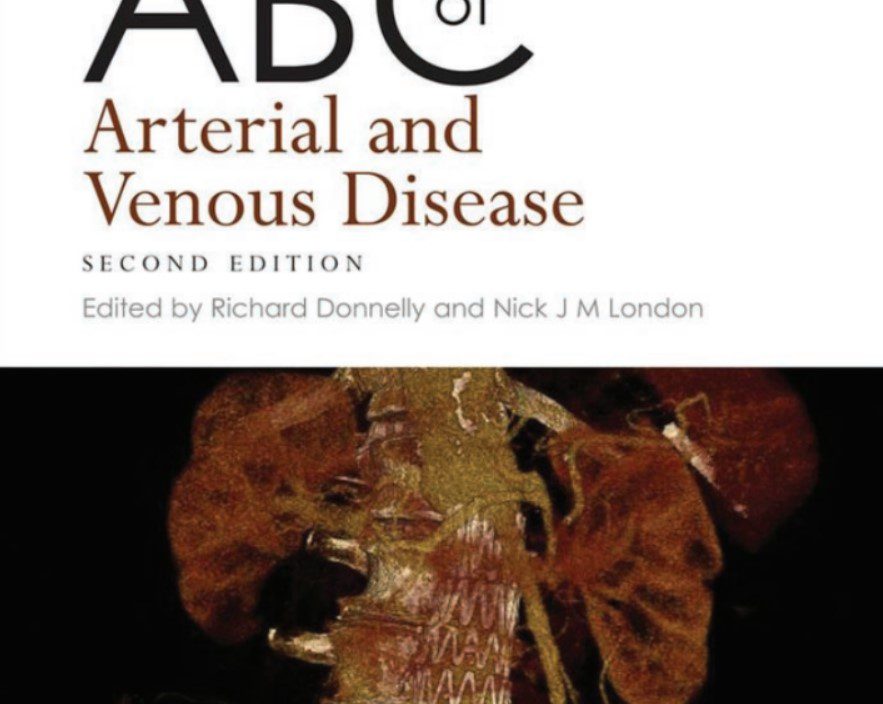
In this blog post, we are going to share a free PDF download of ABC of Arterial and Venous Disease 2nd Edition PDF using direct links. In order to ensure that user-safety is not compromised and you enjoy faster downloads, we have used trusted 3rd-party repository links that are not hosted on our website.
At Medicalstudyzone.com, we take user experience very seriously and thus always strive to improve. We hope that you people find our blog beneficial!
Now before that we move on to sharing the free PDF download of ABC of Arterial and Venous Disease 2nd Edition PDF with you, here are a few important details regarding this book which you might be interested.
The clinical manifestations of arterial and venous disease are often the result of various pathophysiological mechanisms, including atherosclerosis, thrombosis, inflammation, embolism and aneurysm formation.
Recent developments in non-invasive imaging particularly duplex scanning, high resolution CT and magnetic resonance imaging have revolutionised the identifi cation of structural and functional abnormalities in arteries and veins. There have been signifi cant changes in our knowledge of arterial and venous diseases since the last edition of this book in 2000. This revised and updated edition is designed to provide a contemporary and practical description of the techniques used to diagnose, investigate and manage arterial and venous diseases. The authorship refl ects an integrated approach to clinical management involving surgeons, physicians, radiologists and vascular laboratory technicians working closely to achieve optimum outcomes. For example, the approach to carotid disease and renal artery stenosis demonstrates the importance of multidisciplinary input to clinical decision making. This book is targeted primary at the non-specialist and wherever possible, we have tried to ensure that clinical practice recommendations in this book are evidence based. However, clinical trials in patients with arterial and venous disease are relatively limited and there remain gaps in our knowledge. Nevertheless, we hope that this book provides an up-to-date text covering best practice in a rapidly changing and diverse group of common clinical disorders.

Diagnostic and therapeutic decisions in patients with vascular disease are guided primarily by the history and physical examination. However, the accuracy and accessibility of non-invasive investigations have greatly increased due to technological advances in computed tomography (CT) and magnetic resonance (MR) scanning. CT angiography (CTA) and MR angiography (MRA) continue to evolve rapidly, and are best described as ‘minimally invasive’ techniques when used with intravenous (i.v) contrast. This chapter describes the main investigative techniques used in arterial and venous disease. Principles of vascular ultrasound In its simplest form, ultrasound is transmitted as a continuous beam from a probe that contains two piezoelectric crystals. The transmitting crystal produces ultrasound at a fi xed frequency (set by the operator according to the depth of the vessel being examined) whilst the receiving crystal vibrates in response to refl ected waves and produces an output voltage. Conventional B-mode (brightness mode) ultrasonography records the ultrasound waves refl ected from tissue interfaces and a two-dimensional picture is built up according to the refl ective properties of the tissues. Ultrasound signals refl ected off stationary surfaces have the same frequency with which they were transmitted, but the principle underlying Doppler ultrasonography is that signals refl ected from moving objects, e.g. red blood cells, undergo a frequency shift in proportion to the velocity of the target. The output from a continuous- wave Doppler ultrasound is most frequently presented as an audible signal (e.g. a hand-held pencil Doppler, Figure 1.1), so that a sound is heard whenever there is movement of blood in the vessel being examined. With continuous-wave ultrasonography there is little scope for restricting the area of tissue that is being examined because any sound waves that are intercepted by the receiving crystal will produce an output signal. The solution is to use pulsed ultrasound. This enables the investigator to focus on a specifi c tissue plane by transmitting a pulse of ultrasound and closing the receiver except when signals from a predetermined depth are returning. For example, the centre of an artery and the areas close to the vessel wall can be examined in turn.
You might also be interested in:
ABC of Asthma 6th Edition PDF Free Download
ABC of Clinical Electrocardiography 2nd Edition PDF Free Download
ABC of Child Protection 4th Edition PDF Free Download
ABC of Clinical Genetics 3rd Edition PDF Free Download
ABC of Burns PDF Free Download
Download ABC of Arterial and Venous Disease 2nd Edition PDF

Disclaimer:
This site complies with DMCA Digital Copyright Laws.Please bear in mind that we do not own copyrights to this book/software. We are not hosting any copyrighted contents on our servers, it’s a catalog of links that already found on the internet. Medicalstudyzone.com doesn’t have any material hosted on the server of this page, only links to books that are taken from other sites on the web are published and these links are unrelated to the book server. Moreover Medicalstudyzone.com server does not store any type of book,guide, software, or images. No illegal copies are made or any copyright © and / or copyright is damaged or infringed since all material is free on the internet. Check out our DMCA Policy. If you feel that we have violated your copyrights, then please contact us immediately.We’re sharing this with our audience ONLY for educational purpose and we highly encourage our visitors to purchase original licensed software/Books. If someone with copyrights wants us to remove this software/Book, please contact us. immediately.
You may send an email to [email protected] for all DMCA / Removal Requests.You may send an email to [email protected] for all DMCA / Removal Requests.

![ALL MBBS Books PDF 2026 - [First Year to Final Year] Free Download ALL MBBS Books PDF 2022 - [First Year to Final Year] Free Download](https://medicalstudyzone.com/wp-content/uploads/2022/06/ALL-MBBS-Books-PDF-2022-First-Year-to-Final-Year-Free-Download.jpg)


Leave a Reply