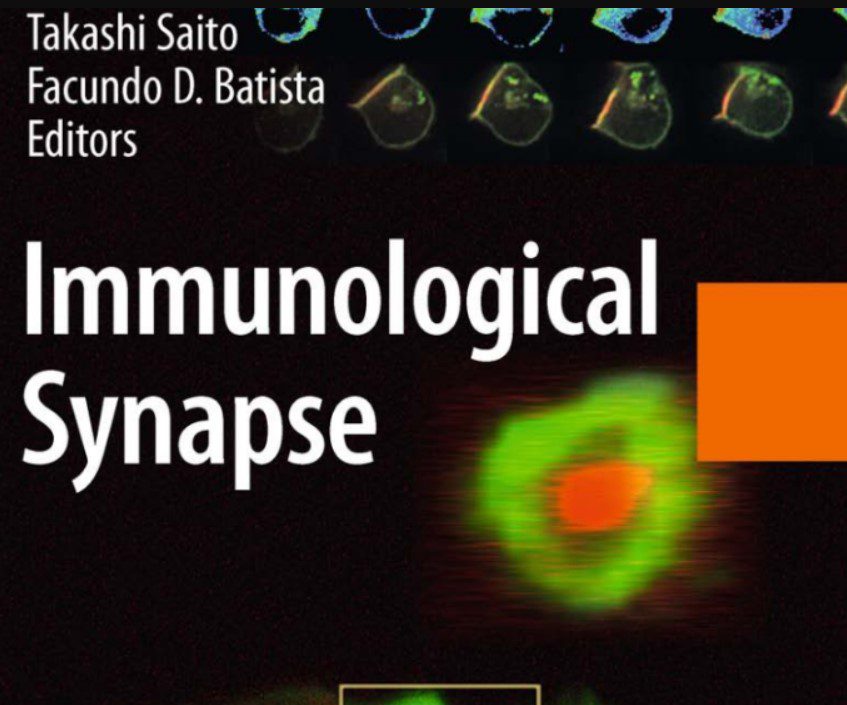
In this blog post, we are going to share a free PDF download of Immunological Synapse PDF using direct links. In order to ensure that user-safety is not compromised and you enjoy faster downloads, we have used trusted 3rd-party repository links that are not hosted on our website.
At Medicalstudyzone.com, we take user experience very seriously and thus always strive to improve. We hope that you people find our blog beneficial!
Now before that we move on to sharing the free PDF download of Immunological Synapse PDF with you, here are a few important details regarding this book which you might be interested.
The proper physiological functioning of most eukaryotic cells requires their assembly into multi-cellular tissues that form organized organ systems. Cells of the immune system develop in bone marrow and lymphoid organs, but as the cells mature they leave these organs and circulate as single cells. Antigen receptors (TCRs) of T cells search for membrane MHC proteins that are bound to peptides derived from infectious pathogens or cellular transformations. The detection of such specific peptide–MHC antigens initiates T cell activation, adhesion, and immune-effectors functions. Studies of normal and transformed T cell lines and of T cells from transgenic mice led to comprehensive understanding of the molecu- lar basis of antigen-receptor recognition and signaling. In spite of these remarkable genetic and biochemical advances, other key physiological mechanisms that parti- cipate in sensing and decoding the immune context to induce the appropriate cellular immune responses remain unresolved.
TCR recognition is tightly regulated to trigger sensitive but balanced T cell responses that result in the effective elimination of the pathogens while minimizing collateral damage to the host. The sensitivity of TCR recognition has to be properly tempered to prevent unintended activation by self-peptide–MHC complexes that cause autoimmune diseases. It is likely that once the TCR is engaged by a peptide– MHC and TCR signaling begins, additional regulatory mechanisms, involving other receptors, would increase the fidelity of the response. Such mechanisms that interpret TCR recognition within physiological settings may provide excellent targets for selective manipulation of immune responses, either enhancing or sup- pressing the immunity. However, the study of T cell activation under physiological conditions faces many technical challenges.

Cloned T cell lines and TCR transgenic mice can address the extremely low frequency of antigen-specific T cells within the circulating mature lymphocytes. Anti-receptor antibodies and soluble purified receptor–ligands are commonly used to selectively engage a particular receptor, even within a polyclonal lymphocyte population, without engaging any of the many other receptors on the immune cells. Such approaches were instrumental for identifying the molecular and functional role of adhesion receptors (LFA-1), co-stimulatory receptors (CD4, CD8, CD28), and inhibitory receptors (CTLA-4), to name just a few. However, these precise
reductionist experiments were not designed to explain the remarkable sensitivity of the T cells and the complex-integrative mechanisms that decode TCR recognition and execute the necessary response. The study of T cell interactions with live APCs, where all activating and inhibitory receptors would be able to bind their physiolo- gical counter-receptors, may be more physiologically appropriate to address these issues.
Molecular imaging, using immunofluorescence microscopy, enabled the study of specific cellular interactions of immune cells. Biophysical considerations predicted that the receptors in T cell that bound their counter-receptors on the membrane of the APC would stabilize the cell contact. Moreover, the extent of receptor clustering at the contact is a quantitative and qualitative measure of receptor–ligand interactions. The first such studies used cloned NKT cells, CTL, and CD4 T cells and demonstrated that these T cells formed antigen-specific cell conjugates with their target cells or antigen-presenting cells (APCs). Immunofluo- rescence microscopy of these cellular conjugates demonstrated remarkable TCR- specific molecular rearrangements at the T–APC cell contact area, known today as the immune synapse. The TCR clustered at the synapse in an antigen-specific manner. Surprisingly, LFA1 and CD4 clustered only when the TCR was engaged, demonstrating that the interactions of LFA1 with ICAM and of CD4 with MHC depended on TCR signals. Thus, engagement of one receptor can affect theresponses of other receptors, even without direct physical intermolecular interac- tions between them.
On the basis of similar consideration, molecular imaging was introduced to identify the intracellular proteins that are involved in signaling and cell adhesion. Immunofluorescence microscopic localization of cytoskeletal proteins identified talin as a key adhesion protein that associates with activated LFA1 and clusters at the synapse. Interestingly, minimal TCR signaling that is not sufficient to produc- tively activate the T cells, or treatment with PMA, is sufficient to cause clustering of talin. Thus, imaging can detect different molecular responses to TCR activation and link them to different cellular outcomes. In another cytoskeletal system, the microtubule network also responded to activation in the T–APC conjugates. The microtubule-organizing center (MTOC) and its associated Golgi apparatus reoriented within minutes in the T cell to face the synapse. Unlike the clustering of talin, MTOC reorientation is induced only in productively activated T cells. Intracellular imaging demonstrated that the role of this early MTOC/Golgi reorientation is to direct the secretion from activated T cells. In CTLs, cytotoxic proteins are directed toward the specific target cells to maximize their effective killing while limiting the damage to innocent bystander cells. In CD4 cells, selective cytokines are directed toward the activating B cells to maximize proliferation and differentiation of antigen-specific B cells that will
produce relevant neutralizing antibodies. Imaging of intracellular proteins detected many of the known proteins that participate in the biochemical pathways of T cell activation at the synapse. Molec-ular imaging can also be used as a screen to identify new proteins that regulate T cell functioning. Such a screen identified PKCy at the synapse, unlike any of the other PKC isoforms. Disruption of the PKCy gene in mice demonstrated that PKCy is essential for T cell proliferation and IL2 production.
The development and use of multilabel three-dimensional deconvolution mi- croscopy was a turning point in the study of the immune synapse. The biggest surprising finding is that two intracellular proteins, talin and PKCy, that cluster at the synapse in response to TCR signaling are spatially segregated at the contact, with PKCy at the center surrounded by a ring of talin. This spatial segregation is mirrored also for receptors, with TCR clustering at the center surrounded by a ring of LFA1. Thus, multiple receptors that are uniformly distributed in the membrane of unbound T cells cluster upon cell conjugation, but rather than just clustering
randomly they formed distinct SupraMolecular Activation Clusters (SMACs).
These molecular structures are not seen when the TCR bound an antagonistic peptide, suggesting that restructuring of the synapse and SMAC formation are essential for T cell functioning. It is important to note that distinct molecular and structural changes take place on both the T cell and the APC side of the synapse. From the time of these initial reports many laboratories extensively studied the structure and function of the synapse in different setups. The use of live cell imaging provided new insights on the kinetic nature of this immune cell junction. Quantitative imaging and mathematical modeling are continually challenging our
views of the synapse. The use of new imaging techniques including TIRF and FRET, which will be discussed in this issue, provide even higher resolution and identify the detailed kinetics of TCR–microcluster formation.
On the other extreme, multiphoton intravital imaging provides new insights on the dynamics of T cell interactions with dendritic cells in live mice. The intravital imaging provides the essential physiological data and context for the T–APC contacts but lacks the molecular resolution provided by other microscopic methods. New imaging tech- niques, that are developed continually, are likely to evolve our understanding of T cell function in health and disease. Interestingly, it appears that human lymphotropic retroviruses like HIV and HTLV1 use the immune synapse to evade elimination. HIV and HTLV1 hijack the endogenous machinery of Golgi/MTOC reorientation and directed secretion to spread stealthily between interacting cells. Such localized viral transmission within confined cell contacts, which are referred to as virological synapses, may limit the access of soluble neutralizing antibodies to the transmitted viruses and may explain how they evade these antibodies. Moreover, these viruses can induce structural alterations of the synapse to reduce anti-viral immunity.
Although the importance of the immune synapse as a dynamic cell junction that regulates intercellular communication and viral transmission is universally recog- nized, many of the quantitative and qualitative details remain unsettled. The plasticity of this cell junction has to accommodate many very different binding partners and control the fate of interacting cells. It is likely that developmental changes in the state of the T cells or the APCs can impact the structure of the synapse. It is hoped that uncovering the ordered molecular rearrangement at the membrane will provide the long sought after clues for the remarkable functional sensitivity of T lymphocytes. These dynamic molecular interactions may eventually explain how engagement of the same TCR can generate different outcomes that resolve the immune challenges. The reviews in this issue present a broad slice of the many diverse topics that are currently intensely debated.
You might also be interested in:
Self Assessment and Review Microbiology Immunology 4th Edition PDF Free Download
Cellular and Molecular Immunology 8th Edition PDF Free Download
Cellular and Molecular Immunology 9th Edition PDF Free Download
How It Works. The Immune System PDF Free Download
Laboratory procedures for full and partial dentures PDF Free Download
Free Books Download PDF: Immunological Synapse

Disclaimer:
This site complies with DMCA Digital Copyright Laws.Please bear in mind that we do not own copyrights to this book/software. We are not hosting any copyrighted contents on our servers, it’s a catalog of links that already found on the internet. Medicalstudyzone.com doesn’t have any material hosted on the server of this page, only links to books that are taken from other sites on the web are published and these links are unrelated to the book server. Moreover Medicalstudyzone.com server does not store any type of book,guide, software, or images. No illegal copies are made or any copyright © and / or copyright is damaged or infringed since all material is free on the internet. Check out our DMCA Policy. If you feel that we have violated your copyrights, then please contact us immediately.We’re sharing this with our audience ONLY for educational purpose and we highly encourage our visitors to purchase original licensed software/Books. If someone with copyrights wants us to remove this software/Book, please contact us. immediately.
You may send an email to [email protected] for all DMCA / Removal Requests.You may send an email to [email protected] for all DMCA / Removal Requests.

![ALL MBBS Books PDF 2026 - [First Year to Final Year] Free Download ALL MBBS Books PDF 2022 - [First Year to Final Year] Free Download](https://medicalstudyzone.com/wp-content/uploads/2022/06/ALL-MBBS-Books-PDF-2022-First-Year-to-Final-Year-Free-Download.jpg)


![All First Aid Book Series PDF 2025 Free Download [36 Books] All First Aid Book Series PDF 2020 Free Download](https://medicalstudyzone.com/wp-content/uploads/2020/07/All-First-Aid-Book-Series-PDF-2020-Free-Download.jpg)

Leave a Reply