ERYTHROPOIESIS
(DEVELOPMENT Of ERYTHROCYTES)
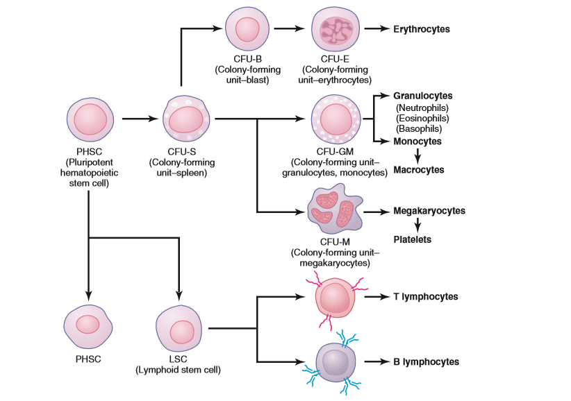
The erythrocytes are derived from the uni-potential progenitor cells called colony forming unit-erythrocyte (CFU-E), which themselves arise from CFU-GEMM. From CFU-E to mature RBC, the erythropoiesis comprises the following stages: Proerythroblast, basophilic erythroblast, polychromatophilic erythroblast, ortho- chromatophilic erythroblast, reticulocyte, and mature erythrocyte.
1.Proerythroblast
The CFU-E differentiates into a proerythroblast which is the earliest recognisable cell of the erythrocyte series. A proerythroblast is a very large cell (15-20 Hm in diameter) having a deeply basophilic cytoplasm. The nucleus, which is spherical and centrally located, shows a uniform chromatin pattern and once or two large nucleoli. Some haemoglobin is present in the cytoplasm, but its amount is too small to be detected by ordinary staining techniques. After undergoing a number of mitotic divisions, the proerythroblasts give rise to basophilic erythroblasts.
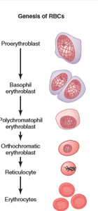
Erythropoiesis
2.Basophilic Erythroblast
This cell is smaller than the proerythroblast and averages 10 pm in diameter. The nucleus shows a coarse network of dense heterochromatin. The Cytoplasm exhibits intense basophilia which indicates a further increase in the number of ribosomes. Haemoglobin increases in amount but is still masked by the basophilia of the cytoplasm.
3.Polychromatophilic Erythroblast
Basophilic erythroblasts undergo a number of mitotic divisions and produce cells in which haemoglobin is present in sufficient quantities to be easily identified in stained smears. With each mitotic division, there is a decrease in the basophilia of cytoplasm and an increase in the quantity of hemoglobin (which is acidophilic) Thus, the cytoplasm of these cells takes varying amounts of acid and basic components of the Wright stain and, therefore, shows mixed colours varying from purplish- blue to a light pink or grey. Hence, these cells are named polychromatophilic erythroblasts.The nucleus of a polychromatophilic erythroblast shows a dense chromatin network with the coarse chromatin masses giving a characteristic checker-board appearance.
4.0rthochromatophilic Erythroblast
Each polychromatophilic erythroblast undergoes a number of mitotic divisions with a gradual decrease in the basophilia and an increase in the amount of haemoglobin. When the cytoplasm becomes as acidophilic as that of a mature erythrocyte, the cells are known as ortho- chromatophilic erythroblasts or normoblasts, An orthochromatophilic erythroblast is only slightly larger than an erythrocyte. Its nucleus is small and condensed and stains deeply basophilic. EM studies reveal that the ribosomes have almost disappeared from the cytoplasm of a normoblast. The Golgi apparatus and mitochondria are also seen to be degenerating. The early orthochromatophilic erythroblasts undergo mitosis actively and eventually reach a stage at which their nuclei become very much shrunken(pyknotic) and no further division is possible. Finally, the nucleus is extruded from each cell.
5.Reticulocyte
The Reticulocytes are immature erythrocytes. Their cytoplasm shows a delicate network(reticulum) when stained by special dyes such as cresyl blue. The reticulocytes contain remnants of the Golgi apparatus, some mitochondria and a few free ribosomes. The RNA of the ribosomes is responsible for the reticulated appearance of the cytoplasm. The reticulocytes are released from the red bone marrow into the circulating blood where they undergo maturation to become red blood cells. The maturation period ranges from 24 to 48 hours. During this period remnants of Golgi apparatus, ribosomes, centrioles and most of the mitochondria are lost to make the cell a mature erythrocyte. Under normal conditions, reticulocytes constitute 0.5 to 2 percent of the circulating erythrocytes.



Leave a Reply