In this blog post, we are going to share a free PDF download of UCSF Abdomen & Pelvis: CT/MR/US 2020 using direct links. In order to ensure that user-safety is not compromised and you enjoy faster downloads, we have used trusted 3rd-party repository links that are not hosted on our website.
At Medicalstudyzone.com, we take user experience very seriously and thus always strive to improve. We hope that you people find our blog beneficial!
Now before that we move on to sharing the free PDF download of UCSF Abdomen & Pelvis: CT/MR/US 2020 with you, here are a few important details regarding this book which you might be interested.
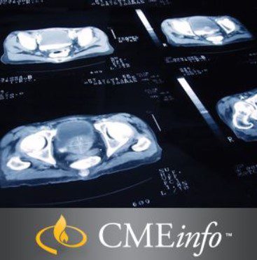
Download UCSF Abdomen & Pelvis: CT/MR/US 2020 Free
UCSF ABDOMEN & PELVIS: CT/MR/US
University of California San Francisco Clinical Update (SA-CME)
Leading radiologists guide you through this updated review of state-of-the-art abdomino-pelvic imaging and interpretation.
Enhance Interpetation Skills
The goal of the UCSF Abdomen & Pelvis: CT/MR/US CME program is to enhance intepretation of body imaging skills and improve clinical practice. This is achieved through a review of state-of-the-art imaging methods and evidence-based practice in CT, MRI and Ultrasound as they pertain to gastrointestinal, genitourinary and gynecologic systems.
Learn how to:
– Apply the ACR recommendations for managing incidental CT-scan findings in the kidney, liver, adrenal gland and pancreas
– Identify indications for the use of different imaging modalities (CT/MRI/US) for various abdomino-pelvic organs and conditions
– Apply practical approaches to diagnosing both benign and malignant diseases of the abdomen and pelvis
– Anticipate potential pitfalls in abdominal-pelvic imaging and interpretation
TOPICS/SPEAKERS
UCSF Abdomen & Pelvis: CT/MR/US Topics
| 1 | Acute Abdominal Emergencies: the Enemy Within – Ronald J. Zagoria, MD |
| 2 | Why We Love to Hate the Spleen: Incidental Findings in Adults and What To Do About Them – John R. Leyendecker, MD |
| 3 | Renal Tumor Ablation: Technique, Results and Follow-up – Ronald J. Zagoria, MD |
| 4 | Transplant Anatomy and Imaging: Kidney, Liver, Pancreas – Marc D. Kohli, MD |
| 5 | Crohn’s Disease of the small bowel – John R. Leyendecker, MD |
| 6 | Non-Interpretive Skills: Informatics and Quality Improvement Basics – Marc D. Kohli, MD |
| 7 | Contrast Media Update: Simpler is Better – Ronald J. Zagoria, MD |
| 8 | Prostate MRI – John R. Leyendecker, MD |
| 9 | Pancreatic Cystic Lesions – Spencer C. Behr, MD |
| 10 | CT Urography: Improved Techniques and Interpretation – Ronald J. Zagoria, MD |
| 11 | Demystifying Vascular Ultrasound (including Carotid Imaging) – Marc D. Kohli, MD |
| 12 | Renal Tumor Imaging – Ronald J. Zagoria, MD |
| 13 | Imaging of the Adrenal Glands – Spencer C. Behr, MD |
| 14 | All You Need to Know About Myllerian Anomalies – Liina Poder, MD |
| 15 | Uterine Fibroid Evaluation: How Imaging Guides Treatment – Marc D. Kohli, MD |
| 16 | Tips and Tricks for Thyroid Imaging and Biopsy – Liina Poder, MD |
| 17 | Non-Obstetrical MRI in Pregnant Patients – Liina Poder, MD |
| 18 | Artifacts and Incidentalomas on FDG PET/CT – Spencer C. Behr, MD |
| 19 | Practical Approach to Adnexal Pathology – Liina Poder, MD |
| 20 | Challenging (and Fun!) Unknown Abdominal/Pelvic Imaging Cases: an Interactive Melee – John R. Leyendecker, MD |
| 21 | Pearls & Pitfalls of Abdominal PET: Non-FDG Avid Tumors – Spencer C. Behr, MD |
| 22 | Multimodality of Endometrial and Cervical Cancer – Liina Poder, MD |
| 23 | Abdominal/Pelvic Complications of Cancer Therapy – John R. Leyendecker, MD |
| 24 | Focal Liver Lesions – Spencer C. Behr, MD |
Learning Objectives
At the completion of this course, you should be able to:
– Discuss management of incidental findings
– Identify indications for the use of different imaging modalities (CT/MRI/US) for various abdomino-pelvic organs and conditions
– Apply practical approaches to diagnosing both benign and malignant diseases of the abdomen and pelvis
– Anticipate potential pitfalls in abdomino-pelvic imaging and interpretation
– Participants of the workshop are expected to learn a) the basic requirements for adequate prostate MR imaging acquisition and b) the use of PIRADS for imaging interpretation
Intended Audience
The activity was designed to help the radiologist determine the indications for the different imaging modalities used for the abdomen and pelvis, as well as indications for secondary imaging studies.
Designation
Series Release: May 1, 2018
Series Expiration: April 30, 2021
You might also be interested in:
UCSF Musculoskeletal MRI 2020 Free Download
UCSF Advances in Internal Medicine 2020 Free Download
UCSF Thoracic Imaging 2020 Free Download
UCSF Breast Imaging 2020 Free Download
UCSF Musculoskeletal Imaging 2020 Free Download
UCSF Abdomen & Pelvis: CT/MR/US 2020 Free Download

Disclaimer:
This site complies with DMCA Digital Copyright Laws.Please bear in mind that we do not own copyrights to this book/software. We are not hosting any copyrighted contents on our servers, it’s a catalog of links that already found on the internet. Medicalstudyzone.com doesn’t have any material hosted on the server of this page, only links to books that are taken from other sites on the web are published and these links are unrelated to the book server. Moreover Medicalstudyzone.com server does not store any type of book,guide, software, or images. No illegal copies are made or any copyright © and / or copyright is damaged or infringed since all material is free on the internet. Check out our DMCA Policy. If you feel that we have violated your copyrights, then please contact us immediately.We’re sharing this with our audience ONLY for educational purpose and we highly encourage our visitors to purchase original licensed software/Books. If someone with copyrights wants us to remove this software/Book, please contact us. immediately.
You may send an email to [email protected] for all DMCA / Removal Requests.You may send an email to [email protected] for all DMCA / Removal Requests.

![ALL MBBS Books PDF 2026 - [First Year to Final Year] Free Download ALL MBBS Books PDF 2022 - [First Year to Final Year] Free Download](https://medicalstudyzone.com/wp-content/uploads/2022/06/ALL-MBBS-Books-PDF-2022-First-Year-to-Final-Year-Free-Download.jpg)


Leave a Reply