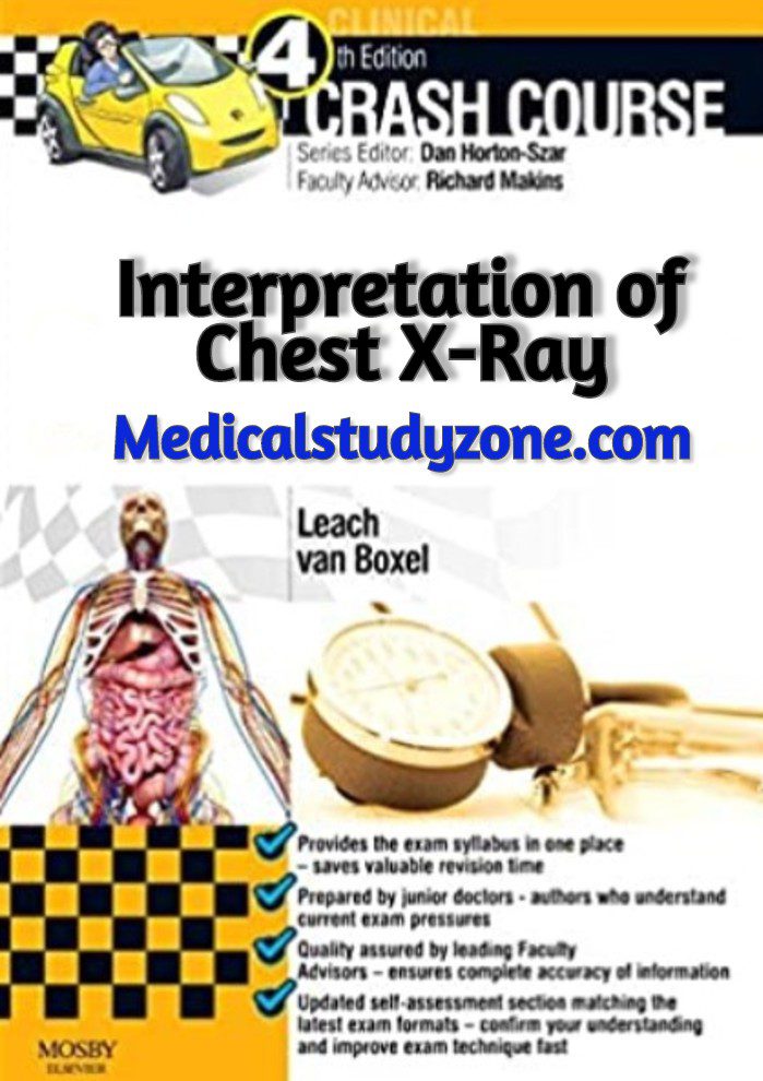
In this blog post, we are going to share a free PDF download of Crash Course Interpretation of Chest X-Ray PDF using direct links. In order to ensure that user-safety is not compromised and you enjoy faster downloads, we have used trusted 3rd-party repository links that are not hosted on our website.
At Medicalstudyzone.com, we take user experience very seriously and thus always strive to improve. We hope that you people find our blog beneficial!

Now before that we move on to sharing the free PDF download of Crash Course Interpretation of Chest X-Ray PDF with you, here are a few important details regarding this book which you might be interested.
To be able to identify the structures or your radio anatomy…To know how a normal chest x-ray appears…To interpret radio pathologic lesion…To put it in practice!
Learning ObjectivesCan anyone in here interpret this chest x-ray for me? (pause)What disease entity is this? Where is this lesion located? front? back? Is this a mass lesion? Consolidation? An atelectasis? How can I interpret this image?I am sure these are some of the few questions which you have in your head right now. Now lets move on to our first case.
This is a chest x-ray in PA view taken from a 36 year old male due to on and off non-productive cough.As you notice in the right paracardiac region, it shows some form of haziness. This was read as a mild inflammatory process. Patient was told take medications but did not complete the regimen.
Patient came back now with a follow-up x-ray done 3 weeks later showed an area of consolidation with air-bronchogram pattern in the right lower lung and right middle lung. Some infiltrates are also seen in the left upper. There is also beginning right pleural effusion.This time, patient took the medication seriously, but refused admission
Somehow the patient agreed to be admitted
Somehow the patient agreed to be admitted. But now presenting with this x-ray!Now we know that effusion progressed. It appears to have a well demarcated borders. We need to know is this fluid free or loculated?Supposing we did not have a previous film for comparison and just be presented with a film that is almost completely opacified hemithorax. It will be hard to know if there is a concomitant mass. Right? So we need to manipulate in order to determine if there is a underlying mass prior to doing a CTT
A right lateral decubitus film was taken showing a free flow of fluid.
Ultrasound showed a 257.23 ml of non loculated pleural effusion.
A lead-lined CTT was inserted with its tip at 5th left posterior rib and its sentinel eye at 6th rib.Now, the radiologist was able to help in the decision making of the AP whether it was safe to proceed with CTT. And was able to give an approximate amount of volume that could be drained.
As a follow-up 2 months after
As a follow-up 2 months after. An impressive clearing of the lungs were seen. So kudos to the clinician and radiologist
BASIC CONCEPTS DENSITIES SOFT TISSUES BONE WATER FAT AIR
Lets go back a little on basics.As you can see, bone or metal appears the most opacified or radioopaque; while air appears the most radiolucent or black. Water and soft tissues for most part, appears as an intermediate density.
BASIC CONCEPTSHere is a schematic of an xray tube. The cathode end which carries the negative charge is accelerated under a vacuum towards the tungsten anode end which is the positive side causing the release of Xray beam. It was named x-ray because during the time of Wilhelm Roentgen in the late 1800’s, such energy form was still unknown, thus the letter “X” in xray. This energy is known to penetrate materials which eventually then made its way in medicine.X-rays.
You might also be interested in:
Crash Course Haematology and Immunology 5th Edition PDF Free Download
Crash Course General Medicine 4th Edition PDF Free Download
Crash Course in Genealogy PDF Free Download
Crash Course Gastrointestinal System 4th Edition PDF Free Download
Crash Course: Foundation Doctor’s Guide to Medicine and Surgery 2nd Edition PDF Free Download
Crash Course Interpretation of Chest X-Ray PDF Free Download

Disclaimer:
This site complies with DMCA Digital Copyright Laws.Please bear in mind that we do not own copyrights to this book/software. We are not hosting any copyrighted contents on our servers, it’s a catalog of links that already found on the internet. Medicalstudyzone.com doesn’t have any material hosted on the server of this page, only links to books that are taken from other sites on the web are published and these links are unrelated to the book server. Moreover Medicalstudyzone.com server does not store any type of book,guide, software, or images. No illegal copies are made or any copyright © and / or copyright is damaged or infringed since all material is free on the internet. Check out our DMCA Policy. If you feel that we have violated your copyrights, then please contact us immediately.We’re sharing this with our audience ONLY for educational purpose and we highly encourage our visitors to purchase original licensed software/Books. If someone with copyrights wants us to remove this software/Book, please contact us. immediately.
You may send an email to [email protected] for all DMCA / Removal Requests.You may send an email to [email protected] for all DMCA / Removal Requests.


![ALL MBBS Books PDF 2026 - [First Year to Final Year] Free Download ALL MBBS Books PDF 2022 - [First Year to Final Year] Free Download](https://medicalstudyzone.com/wp-content/uploads/2022/06/ALL-MBBS-Books-PDF-2022-First-Year-to-Final-Year-Free-Download.jpg)


Leave a Reply