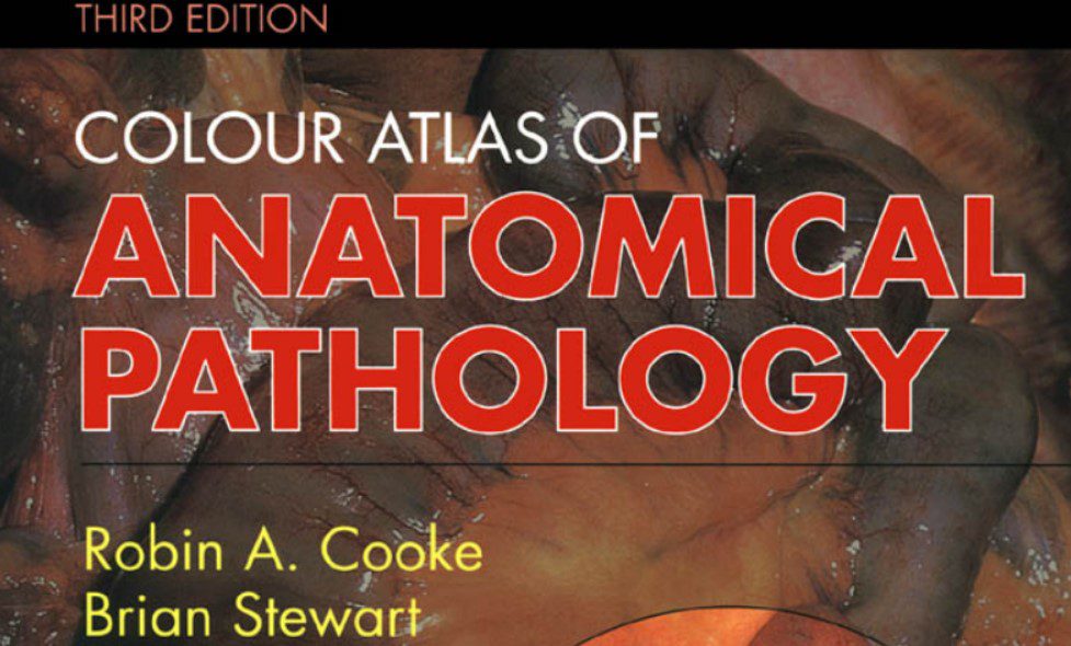
In this blog post, we are going to share a free PDF download of Color Atlas of Anatomical Pathology 3rd Edition PDF using direct links. In order to ensure that user-safety is not compromised and you enjoy faster downloads, we have used trusted 3rd-party repository links that are not hosted on our website.
At Medicalstudyzone.com, we take user experience very seriously and thus always strive to improve. We hope that you people find our blog beneficial!
Now before that we move on to sharing the free PDF download of Color Atlas of Anatomical Pathology 3rd Edition PDF with you, here are a few important details regarding this book which you might be interested.
Color Atlas of Anatomical Pathology – It has often been stated that pathology is primarily a visual discipline. The observation, not always meant as a compliment, has a component of truth, in the sense that a large segment of pathology practice consists in the interpretation of images of one type or another. The magnification range of the images that the pathologist has been asked to evaluate has increased exponentially over the years, from the gross to the microscopic, down to the ultrastructural and cytogenetic; but the basic purpose has remained the same: to extract from the visible structural features all the necessary information in an attempt to ascertain the nature and mechanism of formation of the abnormalities present.
In its beginning, the imagery of pathology was exclusively of a macroscopic nature, and the expertise of the pathologists was once judged on the basis of their acumen in predicting the histology on the basis of the gross appearance of the specimens. Legends were built around this prowess, like the story of Karl Rokitansky looking at a cross-section of a pneumonia and being able to indicate which foci were composed of neutrophils and of histiocytes. A good measure of the importance given at one time to gross pathology is the fact that an integral component of every major department of pathology was the museum, in which a formally appointed curator (none more famous than Thomas Hodgkin) supervised all the activities leading to the selection, processing,
identification and displaying of selected specimens. Color Atlas of Anatomical Pathology

A related activity, which has progressively replaced the actual museums, has been the production and publication of atlases of gross pathology. Some of these were based on the museum pieces and others on fresh specimens, the latter having the obvious advantage of rendering a more faithful representation of the appearance of the ‘live’ lesion. The importance of this material in the teaching of anatomic pathology at both the undergraduate and postgraduate level cannot be overemphasized, particularly at a time when the attitude is gradually taking hold that the examination of a gross specimen is simply the technical step required for the acquisition of the microscopic slides. Nothing can be further from the truth, either in surgical pathology or autopsy pathology. As stated in an editorial appropriately titled ‘In praise of the gross examination’, it is the
gross aspect of the specimen that shows the size, form and nature of the process so that it can be
understood both in a structural sense and in a clinical context. Color Atlas of Anatomical Pathology
For some specimens, a careful gross examination is infinitely superior to the examination of random microscopic sections taken from that specimen. Therefore, books and atlases that display in an optimal fashion the features of these specimens are crucial to the specialty, and I dare say they will remain so long after the genetic background of all human diseases has been determined. Alas, it is not easy to produce such a work. Color Atlas of Anatomical Pathology
First of all, it takes a place that handles a wide range of specimens in order to select those that are most representative of the conditions being depicted. Secondly, it needs a prosecutor who handles those specimens, as Arthur Hertig once put it, ‘with loving care’. Thirdly, it takes a photographer who is not only technically skilled but who also has an aesthetic feeling for those specimens. Unfortunately, these desiderata are rarely found together. Regarding the latter aspect, it has been stated in frustration that ‘[gross] photographs are often not taken, or, when taken, they are not useful because of underexposure, overexposure, inappropriate lighting, poor selection of background, or blood-stained or blood-smeared backgrounds’.I have learned the truth of this statement the hard way when searching the photography archives of one pathology department or another for good pictures for my Surgical Pathology book, only to discard nine out of ten of those pictures because of those very reasons. Color Atlas of Anatomical Pathology
It was therefore with great pleasure, admiration and a touch of envy that I looked at the remarkable
collection of photographs that Dr Robin Cooke has been able to assemble in this Atlas. The specimens are superb, the pictures are technically excellent, the range of diseases they depict is all-encompassing, and the accompanying text is informative while refreshingly succinct. This opus should be of great utility to anybody interested in the pathology of human diseases, whether as companion of a standard textbook or as a stand-alone publication.
You might also be interested in:
Download Color Atlas of Forensic Pathology PDF Free
Applied Respiratory Pathophysiology PDF Free Download
Understanding Pathophysiology PDF Free Download
Robbins Basic Pathology 9th Edition PDF Free Download
Cawson’s Essentials of Oral Pathology and Oral Medicine 8th Edition PDF Free Download
Download Color Atlas of Anatomical Pathology 3rd Edition PDF

Disclaimer:
This site complies with DMCA Digital Copyright Laws.Please bear in mind that we do not own copyrights to this book/software. We are not hosting any copyrighted contents on our servers, it’s a catalog of links that already found on the internet. Medicalstudyzone.com doesn’t have any material hosted on the server of this page, only links to books that are taken from other sites on the web are published and these links are unrelated to the book server. Moreover Medicalstudyzone.com server does not store any type of book,guide, software, or images. No illegal copies are made or any copyright © and / or copyright is damaged or infringed since all material is free on the internet. Check out our DMCA Policy. If you feel that we have violated your copyrights, then please contact us immediately.We’re sharing this with our audience ONLY for educational purpose and we highly encourage our visitors to purchase original licensed software/Books. If someone with copyrights wants us to remove this software/Book, please contact us. immediately.
You may send an email to [email protected] for all DMCA / Removal Requests.You may send an email to [email protected] for all DMCA / Removal Requests.

![ALL MBBS Books PDF 2026 - [First Year to Final Year] Free Download ALL MBBS Books PDF 2022 - [First Year to Final Year] Free Download](https://medicalstudyzone.com/wp-content/uploads/2022/06/ALL-MBBS-Books-PDF-2022-First-Year-to-Final-Year-Free-Download.jpg)


![All First Aid Book Series PDF 2025 Free Download [36 Books] All First Aid Book Series PDF 2020 Free Download](https://medicalstudyzone.com/wp-content/uploads/2020/07/All-First-Aid-Book-Series-PDF-2020-Free-Download.jpg)

Leave a Reply