In this blog post, we are going to share Understanding Basics of Reading Chest X-ray . In order to ensure that user-safety is not compromised and you enjoy faster downloads, we have used trusted 3rd-party repository links that are not hosted on our website.
At Medicalstudyzone.com, we take user experience very seriously and thus always strive to improve. We hope that you people find our blog beneficial!
Now before that we move on to sharing the Understanding Basics of Reading Chest X-ray with you, here are a few important details regarding this book which you might be interested.
X-Ray is a type of radiography and most widely used investigation. It first appears too complicated to read the chest X-Rays because we barely know what lies where and what to make out of it. But the basics of Chest X-Ray here will guide you through various aspects, including Counting ribs, PA vs AP view, Inspiratory vs Expiratory X-Ray, Erect vs Supine, Lucency and Opacity and some common terms like Consolidation and Pleural Effusion.
PA vs AP view
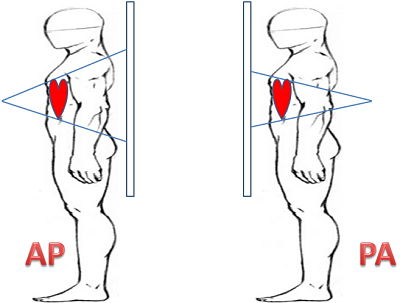
- AP or Anteroposterior view- The view is from front to back.
- PA or Posteroanterior view- The view is from back to front
Difference between PA vs AP view Chest X-Ray
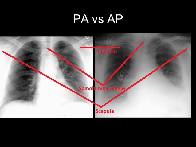
| Features | PA view | AP view |
| Position of clavicle | Oblique | Horizontal |
| Scapula | Away from lung field | Over the lung field |
| Spirolamina angle | Inverted ‘V’ | Not significant |
PA is most common X-Ray done where AP is usually done when the patient cannot stand and X-Ray machine is brought to him on bed and views are taken from anterior to posterior.
The point to add is that there is apparent Cardiomegaly in AP view as compared to PA view because there is a slight magnification of heart since the heart is away from view capturing film.
This can be well understood by the following:-
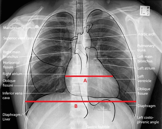
The approach to cardiomegaly on Chest X-Ray is as follows:
- A/B x 100 = cardio ratio
- In PA view, Cardiomegaly when the ratio is more than 50%
- In AP view, Cardiomegaly when the ratio is more than 60%
Erect vs Supine position
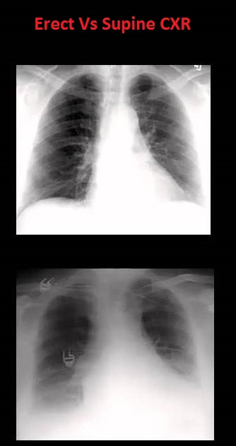
There is fundal view in erect position because all the air in the stomach comes in fundus when the patient is standing.
Inspiratory vs Expiratory
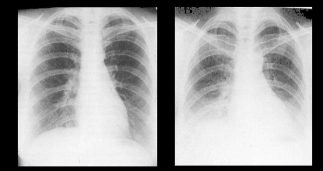
If the anterior end of 6th or 7th rib reaches mid-clavicular line of the diaphragm, it is Inspiratory X-Ray.
Counting Ribs in Chest X-Ray
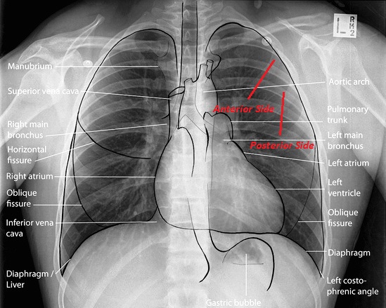
Two points can just help you quickly count ribs from top to bottom:
- The front opaque appearing side of ribs is actually its posterior side.
- Ribs are counted from anterior sides.
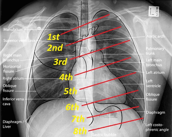
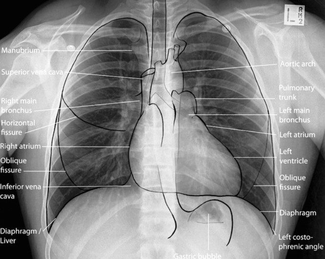
Before we proceed, let us see what structures lie in a normal Chest X-Ray:
The Chest X-Ray is usually divided into three zones as:
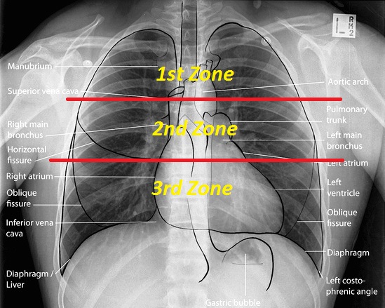
- Up to 2nd rib- First zone
- 2nd to 4th rib- Second zone
- 4th to 6th rib- Third zone
Now let’s proceed to start studying the X-Ray.
Lucency and Opacity in Chest X-Ray
Lucency
Anything that appears dark or black on chest X-Ray is said to be lucent.
- This is because of less density.
- Black color appears because of AIR.
Opacity
Anything that appears light or white on chest X-Ray is said to be Opaque.
- This is because of high density.
- White color appears because of Bones and soft tissues.
Therefore, we can conclude the following easily:-
Increase in lucency:
- Increase in air
- Decrease in soft tissues or absence of bone
Increase in Opacity:
- Increase in soft tissue or abnormal bone
- Decrease in air
The basic approach when seeing a chest X-Ray always sequentially as:-
- Define whether X-Ray is normal or abnormal
- If X-Ray is abnormal, where is this abnormality
- Extent of abnormality
- What is the final diagnosis
Before we proceed to pathological approaches to Chest X-Rays, let’s see what layers the X-Rays hit when they enter the body. Note this strengthens further basics:-
Muscle> Ribs> Pleura> Lung
Talking about when Hyperlucency (increase in blackness) or Hyperopacity (increase in whiteness) occurs:-
Unilateral Lung Hyperopacity
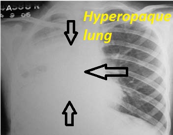
- Consolidation- Replacement of air by something abnormal
- Atelectasis- Collapse of lung resulting in loss of air
Also seen in Plethora, i.e, increase I vascularity.
The differential diagnosis of three important causes if unilateral (one side) opaque thorax are:-
1. Atelectasis- the collapse of the lung

- Displacement of interlobar fissure: because the lobes of lung collapse, the fissures in between the lobes move up or down because of hyperinflation of normal lobe against collapsed lobe. This is the most reliable direct sign of Collapse.
- Mediastinal shift: The structures on mediastinum shift to the side of the collapsed lung
- Crowding of ribs
- Elevation of hemidiaphragm
- Sharp defined margins of opacity
2. Consolidation- replacement of air

- No mediastinal shift
- Ill-defined margins of opacity
- Air bronchogram sign: visualization of air in bronchus surrounded by alveolar opacity
Positive Airbronchogram sign is seen in:
- All except interstitial (viral) pneumonia
- Pulmonary edema (water replace air)
- ARDS (Acute respiratory distress syndrome)
- Goodpasture syndrome (blood)
- HMD (Hyaline membrane disease)
- Pulmonary alveolar proteinosis (macrophages congested in alveoli making a crazy paving pattern)
Air bronchogram sign is NOT seen in:
- Lung abscess
- All except bronchoalveolar carcinoma
3. Pleural effusion-accumulation of fluid
Normally, there is no air in pleura. But effusion in pleura can occur.
- Mediastinal shift: which is on the opposite side, i.e, structures shift to the opposite side of pleural effusion.
Note: Pleural effusion and Haemothorax cannot be differentiated because soft tissue cannot be differentiated on Chest X-Ray.
Unilateral Lung Hyperlucency
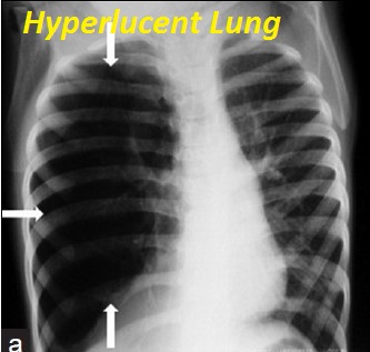
- Rotation: apparent increase in air gap
- Scoliosis
- Mastectomy
- Poland syndrome (absent pectoralis major muscle)
- Airway obstruction
- Large pulmonary embolus
- Pneumothorax
A small mnemonic to quickly grab the names:
POEMS
- P- Poland syndrome/Pneumothorax
- O- Oligemia/Obstruction (like Pulmonary embolism)
- E- Emphysema
- M- Mastectomy/Mucous plug
- S- Swyer’s james syndrome
You might also be interested in:
Download Dorland’s Illustrated Medical Dictionary 33rd Edition PDF Free
Download PATHOMA Lecture Notes 2020 PDF FREE
Download DAMS Handwritten Notes 2020 PDF FREE
Download Marrow Handwritten Notes 2020 PDF FREE [All Subjects]
Download Prepladder Handwritten Notes 2020 PDF FREE
Enjoyed reading? In our upcoming blog of Chest X-Ray, we will be explaining Silhouette sign (where exactly the abnormality is in the lung relating with an intervening border of an organ or its part) and some pathological diseases observed on chest X-Rays like pulmonary embolism, left ventricular failure, bronchogenic carcinoma, bronchiectasis, and lots more. Stay tuned.

Disclaimer:
This site complies with DMCA Digital Copyright Laws.Please bear in mind that we do not own copyrights to this book/software. We are not hosting any copyrighted contents on our servers, it’s a catalog of links that already found on the internet. Medicalstudyzone.com doesn’t have any material hosted on the server of this page, only links to books that are taken from other sites on the web are published and these links are unrelated to the book server. Moreover Medicalstudyzone.com server does not store any type of book,guide, software, or images. No illegal copies are made or any copyright © and / or copyright is damaged or infringed since all material is free on the internet. Check out our DMCA Policy. If you feel that we have violated your copyrights, then please contact us immediately.We’re sharing this with our audience ONLY for educational purpose and we highly encourage our visitors to purchase original licensed software/Books. If someone with copyrights wants us to remove this software/Book, please contact us. immediately.
You may send an email to [email protected] for all DMCA / Removal Requests.You may send an email to [email protected] for all DMCA / Removal Requests.


![FMGE Solutions 7th Edition PDF Free Download [Complete Coloured Book] FMGE Solutions 7th Edition PDF Free Download](https://medicalstudyzone.com/wp-content/uploads/2023/03/FMGE-Solutions-7th-Edition-PDF-Free-Download-150x150.jpg)


Have you uploaded the blog on which explanation about silhouette sign, pulmonary emboli and heart failure are explained?