Determination of various lung volumes and capacities by Spirometry
PRINCIPLE
Dry spirometer is a hand-held spirometer, on which an indicator moves as the air is
exhaled, and only expired air volumes can be measured directly. . .
Wet spirometer consists of a plastic or metal bell within a rectangularor Cylindrical tank, so that air can be added or removed from it. The outer tank contains water and has a tube running through it. It can carry the air above the water level. The floating bottomless bell is inverted over the water containing tank and is connected to an indicator which represents the volume of air. The wet spirometer allows both inspired
and expired gas volumes to be measured.
APPARATUS
Simple spriometer/Expirograph, mouth piece, nose clip and antiseptic solution. Simple Spirometer (student spirometer) (Figure 30)
/
w
Simple spirometer is double walled cylindrical chamber containing water between the M9 cylinders. A” ‘nVElTEU CYlinder which is very light in weight carries a hook ”Y which is attached to balancing weight and to writing pen through a pulley. ““5
The paper on the kyrnograph is calibrated for volume as well as time. There are two
meeds of kymograph slow (60 mmlmin or 1 mm/sec) and fast (1200 mm/min or 20
rum/sec). 10 mm tracing on spiormeter paper which is because of movement of bell is
equd to 300 ml ofchange in volume in the bell (1 mm: 30 ml)
1 Spirometer is connected to the metallic tube with the help of a rubber tube A mouth piece is connected to end of the tube (Figure 31)
4. Kymograph of spirometer works on 220 volt AC.
Floating drum . ’ Oxygen ‘ chamber . u.Recording drum Water Counterbaloncin weight 9 . ._… Mouthpiece Figure 31: basic diagram of simple spirometer
THEORY
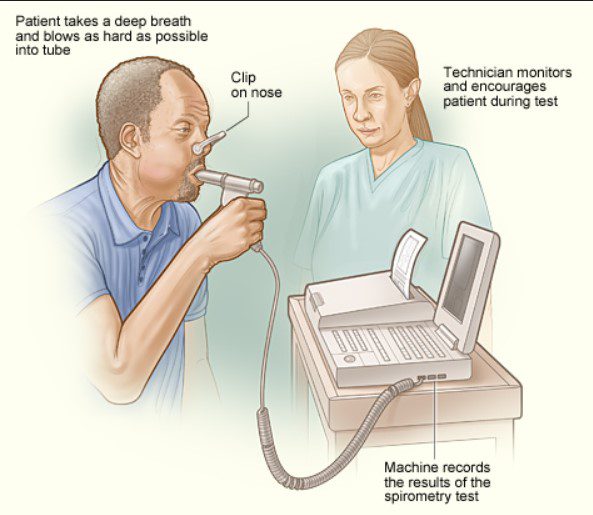
A convenient way of measuring the lung volumes and capacities is by using a Spriometer, and the procedure of recording is called as spinometery. Various lung volumes and capacities are as follows:
1. Tidal Volume (TV): The volume of the air breathed in or out in one breath is called tidal volume. Normal: 500 ml.
2. Inspiratory reserve volume (IRV): It is the maximum volume of the air which can be inspired over and above a normal tidal inspiration with a maximum inspiratory effort. Normal; 2000 to 3200 ml.
1 Expiratory reserve volume (ERV): It is the maximum volume of air which can be expired over and above a normal tidal expiration with a maximum expiratory effort. Normal: 750-1000 mi.
. 4» Residual volume (RV): It is maximal volume of the air which remains in lungs after . a maximum forceful expiration. Normal: 1200 ml.
5Inspiratory capacity (1C): It is the total volume of the air which can be inhaled with
a maximum inspiratory effort. Normal: 2500-3700 ml.
“u
6. Vital capacity (VC): It is the maximum amount of air which can be forcefully exhaled with a maximum expiratory effort after a deep and forceful inspiration.
7. Functional residual capacity (FRC): This is the volume of the air which remains in the lungs at the end of tidal expiration. Normal: 2500 ml. ‘
8. Total lung capacity (TLC): It is the total volume of the air present in the lung at the end of maximal inspiration. Normal: 6 liters.
9. Minute ventilation (MV): It is the volume of the air inspired or expired in one minute. MV = TV + RR. Normal = 500 x 12 = 6000 L/min.
10. Maximum voluntary ventilation (MW) or Maximum ventilation volume: This the volume of the air which is breathed in or out with maximal breathing effort (Deep and fast breathing) in one min. Normal: 90-170 L/min.
11. Pulmonary reserve or Breathing reserve = MW-MV
, MW-MV ‘ Dyspnoeic Index (D1) = _-l7lVV-_ X 100
Normal: 90% < 60% usually associated with dyspnoea.
PROCEDURE
- Fill the distilled water in the spirometer up to 1-2 inches below the upper edge of outer cylinder.
- Clean the mouth piece first with antiseptic solution (1% savalon) and then with water.
- Ask the subject to sit on a stool facing towards the spirometer.
- Give instruction to the subject regarding the experiment so that he will not hesitate at the time of recording.
- Keep the pen of siprometer ready after filling it with ink.
- Fill the spirometer with fresh air by moving its bell up.
- Ask the subject to insert the mouth piece between lips ad teeth. Close the nose With the help of nose clip.
- Subject is asked to breath through the mouth piece in the spirometer quietly. in case of expirograph three way valve is there and with
- the help of it subject breaths from the atmosphere for half to one min. Now connect the subject to spirometer With the help of three
- way valve.
- Start the kymograph, adjust it to move at slow Speed (1 mm/sec). So the Subject inhales a normal breath, and then exhales a normal
- breath of air into the spirometer mouth piece, and the volume is recorded which is known as tidal volume and the
- record is taken for 15 seconds. This graph is used to measure tidal volume and minute
- ventilation. Minute respiratory volume (MRV) is calculated by using the formula:
- MRV = TV x RR (respiratory rate per min) = mls/min.
- Expiratory reserve volume (ERV) is measured by asking the subject exhale forcrbly through the spirometer mouth-piece and ERV is recorded.
Vital capacity (VC) measurement.
Slow speed of kymograph is changed to fast (20 mm/sec). The subject is asked to breathe in and out normally twice or thrice and then he is asked to bend over and exhale all the air possible. Now he is asked to raise in upright position, and inhale as fully as he can. It is very important to strain to inhale maximum amount of air that he can. Asked the subject to quickly insert the mouth piece and exhale as forcefully as he can. Record the results and repeat the test twice. Vital capacity and timed vital capacities are calculated from this spirogram. Vital capacity calculated from the spirogram is also called as forced vital capcmthVC).
Inspiratory reserve volume (IRV) can be calculated using average values obtained from TV, ERV and VC by the equation:
IRV == vc -(Tv + ERV)
Compute the percentage of predicted vital capacity value:
, avera-e measured VC % of predicted VC = predicted value X 100
Same procedures is repeated three times after filling the spirometer With fresh air each time.
For measuring MW spirometer is filled with fresh air and subject is asked to breath in it deep and fast with maximum effort for 15420 seconds. This record is taken with fast Speed (20 mm/sec) of kymograph.
The residual volume (RV) cannot be measured directly, but it can be approximately determined by Using the following factors:
Age Factor
0.250 . $2-23 . 0.305 We
50 69 0.445 \
CALCULATIONS
1.
Tidal Volume Draw two lines in such a way that one line should’touch nmgxggur: number of upper end of tracings and other line should touch maxrmum r 0
lower end of tracings. Measure the vertical distances between two lines. TV = Distance between two lines (mm)x30 = in mi.
10mm vertical distance = 300 ml). _ _ {ung Volume and Capacities Other lung volume and capacrtles are calculated from
the forced vital capacity graph by multiplying the various vertical distances (mm) to 30
mli . . . . FEV1°/o Volume expired in one second of expiration is calculated by consudering the
zero time from the beginning of expiration. (Horizontal 20 mm distance indicates one second).
FEV1 FVE1% = Wx 100
Minute Ventilation (MV): Count inspiratory or expiratory tops for 15 seconds (N) from tidal volume recording. MV = TV x N x 4: L/min
MW Measure the height of all the inspiratory or expiratory tracings for 15 seconds. Add the height of all the tracings (cms).
MVV = Total heght (cm) x 300 (ml) x 4 = L/min. ’: Dyspnoic index is also calculated as discussed in the theory part of practical.
Precautions
VO‘WPWN
Make sure that there should be no water in the rubber t spirometer. Must clean the mouth piece with antiseptic solution.
If. nose clip is being used, clean with alcohol before use. Dispose off used card-board mouth-pieces.
Before recording, practice exhaling through the mouth piece. Take average values (3 readings) of the volumes.
Fill the spirometer with fresh air each time after recording one spirogram.
ube connecting mouth piece to
CLINICAL SIGNIFICANCE OF TIMED VITAL CAPACITY
1. FEV1% is decreased in obstructive lung diseases, e.g. bronchial asthma, but force vital capacity remains normal. _ 2. FEV1% remains normal in case of restrictive lung disease, e.g. pulmonary fibrOSIs, but
FVC decreases. 3. In combined lesion (restrictive + obstructive) FEV1% and FVC both decrease.




Leave a Reply